
Antimicrobial resistance, biofilm formation, and virulence gene profiles of foodborne bacterial isolates from animal-based foods
DOI:
https://doi.org/10.71336/jabs.1587Keywords:
S. aureus, Salmonella spp., antibiotic resistance, virulence, biofilmAbstract
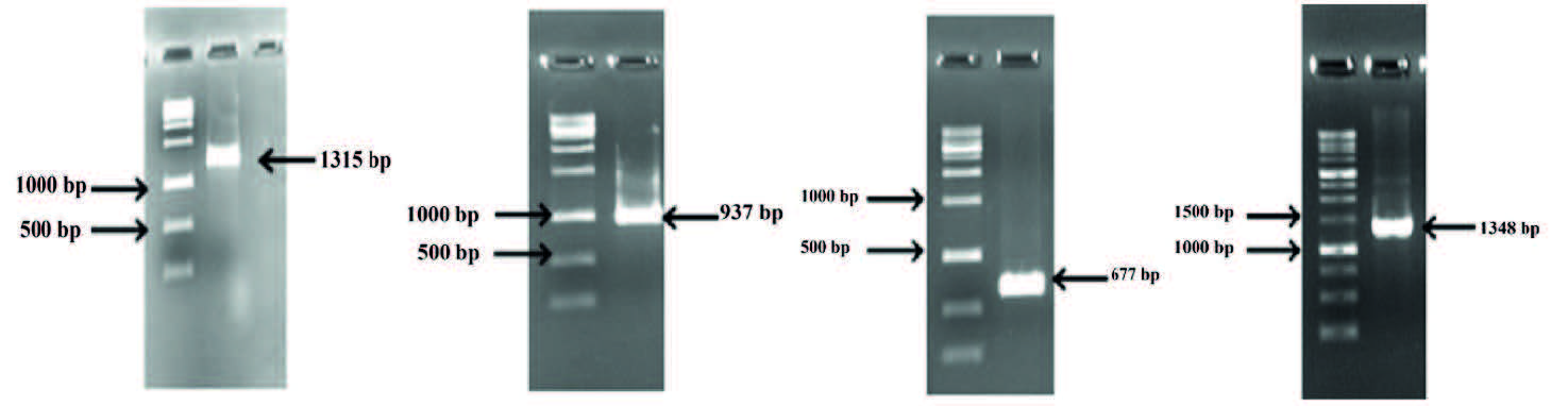
This study aimed to investigate the antibiotic resistance profiles, biofilm-forming capacities, and virulence characteristics of bacteria isolated from 86 animal-based food products, including 25 raw milk, 25 cheese, and 36 raw chicken samples. A total of 40 isolates were identified, comprising 14 Macrococcus caseolyticus, 10 Staphylococcus aureus, 9 Hafnia alvei, and 7 Salmonella spp. isolates. The results demonstrated that slime production was detected in all isolates, while moderate to strong biofilm formation was observed in 90% of the isolates. Among Gram-negative bacteria, resistance to ampicillin was detected in 85.7% of Salmonella spp. and 45.5% of H. Alvei isolates. However, all Gram-negative isolates were susceptible to chloramphenicol, ciprofloxacin, and streptomycin (100%). Regarding Gram-positive bacteria, S. aureus isolates exhibited 20% resistance to tetracycline and erythromycin, whereas M. caseolyticus isolates showed notably higher erythromycin resistance (57.1%). In contrast, all Gram-positive isolates were susceptible to gentamicin and chloramphenicol (100%). Virulence gene analysis revealed that among the S. aureus isolates tested, 30% were positive for icaA and 70% carried These posts must be removed.. Furthermore, among the seven Salmonella spp. isolates analyzed, 85.7% were positive for the mgtC gene, while all isolates (100%) harbored the sopB gene. In conclusion, the detection of S. aureus and Salmonella spp. in animal-based food products, particularly isolates exhibiting strong slime production, biofilmforming ability, and antibiotic resistance, represents a significant public health concern. These findings emphasize the critical importance of maintaining strict hygiene and sanitation practices throughout the production, processing, storage, and distribution stages under appropriate cold chain conditions.
References
[1] Faour-Klingbeil, D., & C. D. Todd, E. (2019): Prevention and Control of Foodborne Diseases in Middle-East North African Countries: Review of National Control Systems. International Journal of Environmental Research and Public Health, 17(1), 70. https://doi.org/10.3390/ijerph17010070. DOI: https://doi.org/10.3390/ijerph17010070
[2] Torgerson, P. R., Devleesschauwer, B., Praet, N., Speybroeck, N., Willingham, A. L., Kasuga, F., Rokni, M. B., Zhou, X. N., Fèvre,
E. M., Sripa, B., Gargouri, N., Fürst, T., Budke, C. M., Carabin, H., Kirk, M. D., Angulo, F. J., Havelaar, A., & de Silva, N. (2015): World Health Organization Estimates of the Global and Regional Disease Burden of 11 Foodborne Parasitic Diseases, 2010: A Data Synthesis. PLoS Medicine, 12(12), e1001920. https://doi.org/10.1371/journal. pmed.1001920. DOI: https://doi.org/10.1371/journal.pmed.1001920
[3] Gallo, M., Ferrara, L., Calogero, A., Montesano, D., & Naviglio,
D. (2020): Relationships between food and diseases: What to know to ensure food safety. Food Research International (Ottawa, Ont.), 137, 109414. https://doi.org/10.1016/j.foodres.2020.109414. DOI: https://doi.org/10.1016/j.foodres.2020.109414
[4] Martinović, T., Andjelković, U., Gajdošik, M. Š., Rešetar, D., & Josić, D. (2016): Foodborne pathogens and their toxins. Journal of Proteomics, 147, 226–235. https://doi.org/10.1016/j. jprot.2016.04.029. DOI: https://doi.org/10.1016/j.jprot.2016.04.029
[5] Chlebicz, A., & Śliżewska, K. (2018): Campylobacteriosis, Salmonellosis, Yersiniosis, and Listeriosis as Zoonotic Foodborne Diseases: A Review. International Journal of environmental research and public health, 15(5), 863. https://doi.org/10.3390/ijerph15050863. DOI: https://doi.org/10.3390/ijerph15050863
[6] Hung, Y. T., Lay, C. J., Wang, C. L., & Koo, M. (2017): Characteristics of nontyphoidal Salmonella gastroenteritis in Taiwanese children: A 9-year period retrospective medical record review. Journal of Infection and Public Health, 10(5), 518–521. https://doi.org/10.1016/j.jiph.2016.09.018. DOI: https://doi.org/10.1016/j.jiph.2016.09.018
[7] Sun, T., Liu, Y., Qin, X., Aspridou, Z., Zheng, J., Wang, X., Li, Z., & Dong, Q. (2021): The Prevalence and Epidemiology of Salmonella in Retail Raw Poultry Meat in China: A Systematic Review and Meta-Analysis. Foods (Basel, Switzerland), 10(11), 2757. https://doi.org/10.3390/foods10112757. DOI: https://doi.org/10.3390/foods10112757
[8] Whiley, H., & Ross, K. (2015): Salmonella and eggs: from production to plate. International Journal of Environmental Research and Public Health, 12(3), 2543–2556. https://doi.org/10.3390/ijerph120302543. DOI: https://doi.org/10.3390/ijerph120302543
[9] Wessels, K., Rip, D., & Gouws, P. (2021): Salmonella in Chicken Meat: Consumption, Outbreaks, Characteristics, Current Control Methods and the Potential of Bacteriophage Use. Foods (Basel, Switzerland), 10(8), 1742. https://doi.org/10.3390/foods10081742. DOI: https://doi.org/10.3390/foods10081742
[10] Yeh, Y., Purushothaman, P., Gupta, N., Ragnone, M., Verma, S. C., & de Mello, A. S. (2017): Bacteriophage application on red meats and poultry: Effects on Salmonella population in final ground products. Meat Science, 127, 30–34. https://doi.org/10.1016/j. meatsci.2017.01.001. DOI: https://doi.org/10.1016/j.meatsci.2017.01.001
[11] Kore, K., Asrade, B., Demissie, K., & Aragaw, K. (2017): Characterization of Salmonella isolated from apparently healthy slaughtered cattle and retail beef in Hawassa, southern Ethiopia. Preventive Veterinary Medicine, 147, 11–16. https://doi. org/10.1016/j.prevetmed.2017.08.018 DOI: https://doi.org/10.1016/j.prevetmed.2017.08.018
[12] Ferrari, R. G., Rosario, D. K. A., Cunha-Neto, A., Mano, S. B., Figueiredo, E. E. S., & Conte-Junior, C. A. (2019): Worldwide Epidemiology of Salmonella Serovars in Animal-Based Foods: a Meta-analysis. Applied and Environmental Microbiology, 85(14), e00591-19. https://doi.org/10.1128/AEM.00591-19. DOI: https://doi.org/10.1128/AEM.00591-19
[13] Asal, C. (2021): Salmonella Bakterisinin Gıdalarda Varlığı. [The Presence of Salmonella Bacteria in Food]. Samsun Sağlık Bilimleri Dergisi, 6(1), 28-34. https://doi.org/10.47115/jshs.695685. DOI: https://doi.org/10.47115/jshs.695685
[14] Atasever, M. (2017): Kanatlı Etlerinde Salmonella Riski. [Salmonella Risk in Poultry Meat]. Atatürk Üniversitesi Veteriner Bilimleri Dergisi, 12(1), 90-98. https://doi.org/10.17094/ataunivbd.309781 DOI: https://doi.org/10.17094/ataunivbd.309781
[15] Özdemir, H., & Keyvan, E. (2016): Isolation and characterization of Staphylococcus aureus strains isolated from beef, sheep and chicken meat. Ankara Üniversitesi Veteriner Fakültesi Dergisi, 63(4), 333-
338. https://doi.org/10.1501/Vetfak_0000002749. DOI: https://doi.org/10.1501/Vetfak_0000002749
[16] Hennekinne, J. A., Ostyn, A., Guillier, F., Herbin, S., Prufer, A. L., & Dragacci, S. (2010): How should staphylococcal food poisoning outbreaks be characterized?. Toxins, 2(8), 2106–2116. https://doi. org/10.3390/toxins2082106. DOI: https://doi.org/10.3390/toxins2082106
[17] Pinchuk, I. V., Beswick, E. J., & Reyes, V. E. (2010): Staphylococcal enterotoxins. Toxins, 2(8), 2177–2197. https://doi.org/10.3390/toxins2082177. DOI: https://doi.org/10.3390/toxins2082177
[18] Mashouf, R. Y., Hosseini, S. M., Mousavi, S. M., & Arabestani,
M. R. (2015): Prevalence of Enterotoxin Genes and Antibacterial Susceptibility Pattern of Staphylococcus aureus Strains Isolated from Animal Originated Foods in West of Iran. Oman Medical Journal, 30(4), 283–290. https://doi.org/10.5001/omj.2015.56. DOI: https://doi.org/10.5001/omj.2015.56
[19] Argudín, M. Á., Mendoza, M. C., & Rodicio, M. R. (2010): Food poisoning and Staphylococcus aureus enterotoxins. Toxins, 2(7), 1751–1773. https://doi.org/10.3390/toxins2071751. DOI: https://doi.org/10.3390/toxins2071751
[20] Moura, E. O., Rangel, A. H., Macêdo, C. S., Urbano, S. A., Novaes, L. P., & Lima, D. M. (2019): Enterotoxin-encoding genes in Staphylococcus aureus from buffalo milk. Pesquisa Veterinária Brasileira, 39(08), 587-591. DOI: https://doi.org/10.1590/1678-5150-pvb-6011
[21] Temel, A., Eraç, B. (2018): Bakteriyel biyofilmler: Saptama yöntemleri ve antibiyotik direncindeki rolü. [Bacterial biofilms: Detection methods and their role in antibiotic resistance]. Türk Mikrobiyoloji Cemiyeti Dergisi 48(1),1-13. DOI: https://doi.org/10.5222/TMCD.2018.001
[22] Kartal, M. O., Baran Ekinci, M., & Poyraz, B. (2021): Biyofilm Yapısı ve Önlenmesi. [Biofilm Structure and Prevention]. Akademik Gıda, 19(3), 353-363. https://doi.org/10.24323/akademik-gida.1011231 DOI: https://doi.org/10.24323/akademik-gida.1011231
[23] Demirtürk, N., Demirdal, T. (2004): Antibiyotiklerde direnç sorunu.[The problem of antibiotic resistance]. Kocatepe Tıp Dergisi, 5:(2).
[24] Ünal Turhan, E., & Erginkaya, Z. (2019): Bakteriyel Biyofilmlerdeki Antimikrobiyel Direnç Mekanizması. [Mechanisms of Antimicrobial Resistance in Bacterial Biofilms]. Akademik Gıda, 17(1), 131-139. https://doi.org/10.24323/akademik-gida.544978 DOI: https://doi.org/10.24323/akademik-gida.544978
[25] Donlan R. M. (2002): Biofilms: microbial life on surfaces. Emerging Infectious Diseases, 8(9), 881–890. https://doi.org/10.3201/eid0809.020063. DOI: https://doi.org/10.3201/eid0809.020063
[26] Bilgehan, H. (1994): Klinik Mikrobiyoloji Özel Bakteriyoloji ve Bakteri Enfeksiyonları, [Clinical Microbiology: Special Topics in Bacteriology and Bacterial Infections]. Bornova: Barış Yayınları,
188- 211.
[27] Heperkan, D. (2016): Temel Gıda Mikrobiyolojisi, [Fundamentals of Food Microbiology]. Ankara: Nobel Yayınları, 323-342.
[28] Anonymous.(1981): Çiğ Süt [Raw Milk] Türk Standartları Enstitüsü, TS 1018.
[29] Anonymous.(1998):TS 3135, ISO 3100-1 Et ve et mamulleri Numune alma ve analiz numunelerinin hazırlanması bölüm 1-Numune alma.[Meat and meat products Sampling and preparation of samples for analysis, Part 1-Sampling].Türk Standartları Enstitüsü, Ankara.
[30] CLSI. (2018): Clinical and Laboratory Institute. Performance Standards for Antimicrobial Susceptibility Testing; Twentyfifth Informational Supplement. Document M100-S23. Clinical and Laboratory Standards Institute.
[31] Christensen, G. D., Simpson, W. A., Younger, J. J., Baddour, L. M., Barrett, F. F., Melton, D. M., & Beachey, E. H. (1985): Adherence of coagulase-negative staphylococci to plastic tissue culture plates: a quantitative model for the adherence of staphylococci to medical devices. Journal of Clinical Microbiology, 22(6), 996–1006. https://doi.org/10.1128/jcm.22.6.996-1006.1985. DOI: https://doi.org/10.1128/jcm.22.6.996-1006.1985
[32] Chusri, S., Phatthalung, P. N., & Voravuthikunchai, S. P. (2012): Anti-biofilm activity of Quercus infectoria G. Olivier against methicillin-resistant Staphylococcus aureus. Letters in Applied Microbiology, 54(6), 511–517.https://doi.org/10.1111/j.1472-765X.2012.03236.x. DOI: https://doi.org/10.1111/j.1472-765X.2012.03236.x
[33] Kaynarca, S., Türkyılmaz, S. (2020): Methicillin resistance and slime positivity of Staphylococci isolated from bovine mastitis. Kafkas Üniversitesi Veteriner Fakültesi Dergisi (16):567-572.
[34] https://www.sigmaaldrich.com/TR/tr/sds/roche/11796828001 Erişim: 19 Nisan 2026 [Online].
[35] Suvajdžić, B., Teodorović, V., Vasilev, D., Karabasil, N., Dimitrijević, M., Đorđević, J. & Katić, V. (2017): Detection of icaA and icaD genes of Staphylococcus aureus isolated in cases of bovine mastitis in the Republic of Serbia. Acta Veterinaria, 67(2),
168- 177. https://doi.org/10.1515/acve-2017-0015. DOI: https://doi.org/10.1515/acve-2017-0015
[36] Kumar, J. D., Negi, Y. K., Gaur, A., & Khanna, D. (2009): Detection of virulence genes in Staphylococcus aureus isolated from paper currency. International Journal of Infectious Diseases: IJID: official publication of the International Society for Infectious Diseases, 13(6), e450–e455. https://doi.org/10.1016/j.ijid.2009.02.020 DOI: https://doi.org/10.1016/j.ijid.2009.02.020
[37] Soto, S. M., Rodríguez, I., Rodicio, M. R., Vila, J., & Mendoza,
M. C. (2006): Detection of virulence determinants in clinical strains of Salmonella enterica serovar Enteritidis and mapping on macrorestriction profiles. Journal of Medical Microbiology, 55(Pt 4), 365–373. https://doi.org/10.1099/jmm.0.46257-0. DOI: https://doi.org/10.1099/jmm.0.46257-0
[38] Osman, K. M., Marouf, S. H., Zolnikov, T. R., & AlAtfeehy, N.(2014): Isolation and characterization of Salmonella enterica in day-old ducklings in Egypt. Pathogens and Global Health, 108(1), 37–48. https://doi.org/10.1179/2047773213Y.0000000118. DOI: https://doi.org/10.1179/2047773213Y.0000000118
[39] Normanno, G., La Salandra, G., Dambrosio, A., Quaglia, N. C., Corrente, M., Parisi, A., Santagada, G., Firinu, A., Crisetti, E., & Celano, G. V. (2007): Occurrence, characterization and antimicrobial resistance of enterotoxigenic Staphylococcus aureus isolated from meat and dairy products. International Journal of Food Microbiology, 115(3), 290–296. https://doi.org/10.1016/j. ijfoodmicro.2006.10.049. DOI: https://doi.org/10.1016/j.ijfoodmicro.2006.10.049
[40] De Luca, G., Zanetti, F., & Stampi, S. (1997): Staphylococcus aureus in dairy products in the Bologna area. International Journal of Food Microbiology, 35(3), 267–270. https://doi.org/10.1016/s0168-1605(96)01229-9. DOI: https://doi.org/10.1016/S0168-1605(96)01229-9
[41] Guven, K., Mutlu, M.B., Gulbandilar, A., & Çakır, P. (2010): Occurrence and Characterization of Staphylococcus aureus Isolated From Meat and Dairy Products Consumed in Turkey. Journal of Food Safety, 30, 196-212. DOI: https://doi.org/10.1111/j.1745-4565.2009.00200.x
[42] Molla, B., & Mesfin, A. (2003): A survey of Salmonella contamination in chicken carcass and giblets in Central Ethiopia.
[43] Aarestrup, F. M., Hendriksen, R. S., Lockett, J., Gay, K., Teates, K., McDermott, P. F., White, D. G., Hasman, H., Sørensen, G., Bangtrakulnonth, A., Pornreongwong, S., Pulsrikarn, C., Angulo, F. J., & Gerner-Smidt, P. (2007): International spread of multidrug-resistant Salmonella Schwarzengrund in food products. Emerging Infectious Diseases, 13(5), 726–731. https://doi.org/10.3201/eid1305.061489. DOI: https://doi.org/10.3201/eid1305.061489
[44] Cetinkaya, F., Cibik, R., Soyutemiz, G. E., Ozakin, C., Kayali, R., ve Levent, B. (2008): Shigella and Salmonella contamination in various foodstuffs in Turkey. Food Control, 19(11), 1059-1063. DOI: https://doi.org/10.1016/j.foodcont.2007.11.004
[45] Erdoğmuş, S. F., & Konak, S. (2020): Bazı Antibiyotiklerin Biyofilm Oluşturan Stafilokok İzolatları Üzerindeki Etkilerinin Değerlendirilmesi. [Evaluation of the Effects of Certain Antibiotics on Biofilm-Forming Staphylococcus Isolates]. Journal of the Institute of Science and Technology, 10(2), 838-845. https://doi. org/10.21597/jist.615352 DOI: https://doi.org/10.21597/jist.615352
[46] Gündoğan, N., Ataol, Ö. (2012): Et örneklerinden izole edilen Staphylococcus aureus ve koagülaz negatif stafilokok’ların biyofilm üretimi ve DNaz aktivitelerinin belirlenmesi.[ Determination of biofilm formation and DNase activity in Staphylococcus aureus and coagulase-negative staphylococci isolated from meat samples] Türk Hijyen ve Deneysel Biyoloji Dergisi, 69(3), 135-142.
[47] Ben Abdallah, F., Lagha, R., Said, K., Kallel, H., & Gharbi, J.(2014): Detection of cell surface hydrophobicity, biofilm and fimbirae genes in Salmonella isolated from Tunisian clinical and poultry meat. Iranian Journal of Public Health, 43(4), 423–431.
[48] Lin, Q., Sun, H., Yao, K., Cai, J., Ren, Y., & Chi, Y. (2019): The Prevalence, Antibiotic Resistance and Biofilm Formation of Staphylococcus aureus in Bulk Ready-To-Eat Foods. Biomolecules, 9(10), 524. https://doi.org/10.3390/biom9100524. DOI: https://doi.org/10.3390/biom9100524
[49] Xing, X., Li, G., Zhang, W., Wang, X., Xia, X., Yang, B., & Meng,
J. (2014): Prevalence, antimicrobial susceptibility, and enterotoxin gene detection of Staphylococcus aureus isolates in ready-to-eat foods in Shaanxi, People’s Republic of China. Journal of Food Protection, 77(2), 331–334. https://doi.org/10.4315/0362-028X.JFP-13-301. DOI: https://doi.org/10.4315/0362-028X.JFP-13-301
[50] Wang, H., Ye, K., Wei, X., Cao, J., Xu, X., Zhou, G. (2013): Occurrence, antimicrobial resistance and biofilm formation of Salmonella isolates from a chicken slaughter plant in China. Food Control, 33(2), 378-384. DOI: https://doi.org/10.1016/j.foodcont.2013.03.030
[51] Farahani, R. K., Ehsani, P., Ebrahimi-Rad, M., ve Khaledi, A.(2018): Molecular Detection, Virulence Genes, Biofilm Formation, and Antibiotic Resistance of Salmonella enterica serotype enteritidis Isolated from Poultry and Clinical Samples. Jundishapur Journal of Microbiology. 11. 10.5812/jjm.69504.
[52] Sepin, Ö., Pamuk, Ş. (2021): Afyonkarahisar’da Satışa Sunulan Tavuk İç Organlarından Salmonella spp. ve Listeria monocytogenes’in İmmonomagnetik Seperasyon Yöntemi ile İzolasyonu ve İzolatların Antibiyotik Dirençliliğinin Belirlenmesi.[ Isolation of Salmonella spp. and Listeria monocytogenes from Chicken Offal Sold in Afyonkarahisar Using the Immunomagnetic Separation Method and Determination of Antibiotic Resistance in the Isolates] KocatepeVeterinaryJournal, 14(2),201-209. https://doi. org/10.30607/kvj.839629 DOI: https://doi.org/10.30607/kvj.839629
[53] Oliver, J. P., Gooch, C. A., Lansing, S., Schueler, J., Hurst, J. J., Sassoubre, L., Crossette, E. M., & Aga, D. S. (2020): Invited review: Fate of antibiotic residues, antibiotic-resistant bacteria, and antibiotic resistance genes in US dairy manure management systems. Journal of Dairy Science, 103(2), 1051–1071. https://doi. org/10.3168/jds.2019-16778. DOI: https://doi.org/10.3168/jds.2019-16778
[54] Ağay, Z., Kimiran, A. (2017): Farklı Kaynaklardan İzole Edilen Salmonella Suşlarının Bazı Virülans Faktörlerinin Belirlenmesi.[Identification of Certain Virulence Factors in Salmonella Strains Isolated from Different Sources]. Süleyman Demirel Üniversitesi Fen Bilimleri Enstitüsü Dergisi, 21(3), 910-916. DOI: https://doi.org/10.19113/sdufbed.91790
[55] Gargano, V., Gambino, D., Migliore, S., Vitale, M., Sciortino, S., Costa, A., & Vicari, D. (2021): Can Human Handling Increase the Presence of Multidrug Resistance (MDR) in Salmonella spp. Isolated from Food Sources?. Microorganisms, 9(10), 2018. https://doi.org/10.3390/microorganisms9102018. DOI: https://doi.org/10.3390/microorganisms9102018
[56] Babacan, O., Karadeniz, H. (2019): Çiğ tavuk etlerinden izole edilen Salmonella spp. suşlarının antibiyotik duyarlılıklarının araştırılması. Journal of Turkish Veterinary Medical Society, 90(2),
105- 114. https://doi.org/10.33188/vetheder.497569 DOI: https://doi.org/10.33188/vetheder.497569
[57] Schleifer, K. H., Kilpper-Bälz, R., Fischer, U., Faller, A. and Endl,
J. (1982): Identification of Micrococcus candidus ATCC 14852 as a strain of Staphylococcus epidermidis and of Micrococcus caseolyticus ATCC 13548 and Micrococcus varians ATCC 29750 as members of a new species, Staphylococcus caseolytic. International Journal of Systematic and Evolutionary Microbiology, 32(1), 15-20. DOI: https://doi.org/10.1099/00207713-32-1-15
[58] MacFadyen, A. C., Fisher, E. A., Costa, B., Cullen, C., & Paterson, G. K. (2018): Genome analysis of methicillin resistance in Macrococcus caseolyticus from dairy cattle in England and Wales. Microbial Genomics, 4(8), e000191. https://doi.org/10.1099/mgen.0.000191 DOI: https://doi.org/10.1099/mgen.0.000191
[59] Blaiotta, G., Pennacchia, C., Villani, F., Ricciardi, A., Tofalo, R., & Parente, E. (2004): Diversity and dynamics of communities of coagulase-negative staphylococci in traditional fermented sausages. Journal of Applied Microbiology, 97(2), 271–284. https://doi. org/10.1111/j.1365-2672.2004.02298.x. DOI: https://doi.org/10.1111/j.1365-2672.2004.02298.x
[60] Resende, J. A., Fontes, C. O., Ferreira-Machado, A. B., Nascimento,
T. C., Silva, V. L., & Diniz, C. G. (2018): Antimicrobial-Resistance Genetic Markers in Potentially Pathogenic Gram Positive Cocci Isolated from Brazilian Soft Cheese. Journal of Food Science, 83(2), 377–385. https://doi.org/10.1111/1750-3841.14019. DOI: https://doi.org/10.1111/1750-3841.14019
[61] Dugat-Bony, E., Sarthou, A. S., Perello, M. C., de Revel, G., Bonnarme, P., & Helinck, S. (2016): The effect of reduced sodium chloride content on the microbiological and biochemical properties of a soft surface-ripened cheese. Journal of Dairy Science, 99(4), 2502–2511. https://doi.org/10.3168/jds.2015-10502. DOI: https://doi.org/10.3168/jds.2015-10502
[62] Szweda, P., Schielmann, M., Milewski, S., Frankowska, A., & Jakubczak, A. (2012): Biofilm production and presence of ica and bap genes in Staphylococcus aureus strains isolated from cows with mastitis in the eastern Poland. Polish Journal of Microbiology, 61(1), 65–69. DOI: https://doi.org/10.33073/pjm-2012-009
[63] Castelani, L., Pilon, L. E., Martins, T., Pozzi, C. R., & Arcaro, J.
R. (2015): Investigation of biofilm production and icaA and icaD genes in Staphylococcus aureus isolated from heifers and cows with mastitis. Animal Science Journal = Nihon Chikusan Gakkaiho, 86(3), 340–344. https://doi.org/10.1111/asj.12284. DOI: https://doi.org/10.1111/asj.12284
[64] Bhatia, A., Zahoor, S., (2007): Staphylococcus aureus Enterotoxins: A Review. Journal of Clinical and Diagnostic Research, 1(2): 188- 97.
[65] Günthard, H., & Pennekamp, A. (1996): Clinical significance of extraintestinal Hafnia alvei isolates from 61 patients and review of the literature. Clinical Infectious Diseases: an Official Publication of the Infectious Diseases Society of America, 22(6), 1040–1045. https://doi.org/10.1093/clinids/22.6.1040. DOI: https://doi.org/10.1093/clinids/22.6.1040
Downloads
Published
How to Cite
Issue
Section
License
Copyright (c) 2026 Journal of Applied Biological Sciences

This work is licensed under a Creative Commons Attribution 4.0 International License.
